Undergraduate Research Center
International Conference of Undergraduate Research (ICUR)
ICUR, the International Conference of Undergraduate Research, is the annual interdisciplinary academic conference that showcases the best in undergraduate research from around the world through our network of participating institutions.
ICUR connects MTSU students with an international audience. Students present their research to a global academic community, enhancing cross-cultural communication skills, expanding their academic perspectives, and increasing their visibility on the world stage.
This two-day academic conference showcases excellence in undergraduate research from around the world. This is a unique opportunity to present your research to interdisciplinary audiences across international borders in real-time, video-linked sessions from their home campus.
MTSU’s Undergraduate Research Center (URC) assists select students in applying for and participating in ICUR.
NEW: ICUR Virtual at MTSU
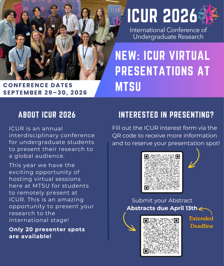
This year, MTSU is proud to host virtual presentation sessions that allow students to participate in the International Conference of Undergraduate Research from campus. Through these sessions, students can remotely present their work to an international audience while benefiting from local support and a professional presentation environment.
This opportunity offers a unique platform to share your research on a global stage, engage with scholars from around the world, and gain valuable experience presenting in an international academic setting. Students selected for these virtual sessions will represent MTSU while contributing to a global exchange of ideas that defines undergraduate research at its highest level.
ICUR 2025
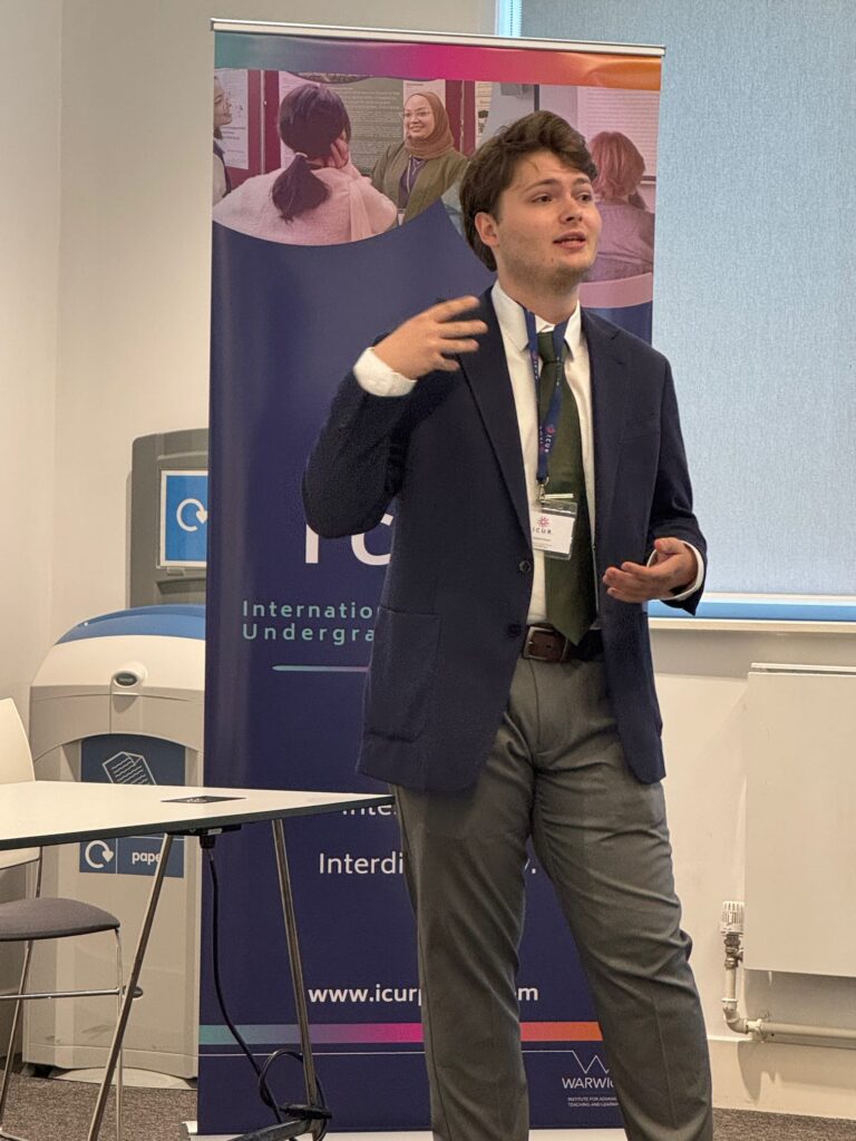
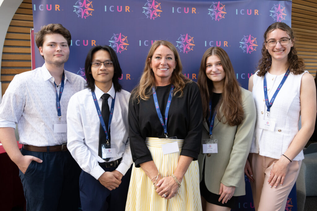
Representing MTSU at the International Conference on Undergraduate Research (ICUR) in England, Andrew Hetrick, Muny Chet, Emily Callison, and Ariel Nicastro showcased not only shared their cutting-edge research but also took on a key leadership role, mentoring peers and facilitating presentations. They gained invaluable experience in planning, teamwork, and cross-cultural collaboration, skills that will continue to shape their academic and professional journey. This trip truly exemplifies the power of undergraduate research and collaboration.


Apply to ICUR
Student researchers are encouraged to apply for the International Conference on Undergraduate Research for global exposure, interdisciplinary exploration, networking opportunities, comprehensive support, international collaboration, and more! We also invite faculty, mentors, and the undergraduate research community to participate in ICUR to advocate for and celebrate this community and to further their professional development through various mentoring sessions
ICUR 2026 applications have closed, but stay tuned for next year’s cycle!
Abstract Selection Process
Being accepted and attending ICUR is an impressive accomplishment and an opportunity for you to disseminate and celebrate your academic achievements. ICUR accepts abstract submissions from across a variety of disciplines. Faculty mentors are expected to take an active role in helping students plan, prepare, and practice their presentations.
The deadline to apply to ICUR has passed for the 2026 conference, but information regarding next year will be posted here too!

Gabriel Greene

Delaney Reynolds

Isaac Robichau

Madison Yahn
MTSU’s 2025 Selected ICUR Participants

Emily Callison
Quantifying the Growth Rate of Cryptococcus neoformans in Macrophage-Like Cells Using Live Cell Imaging
Faculty Mentor: Dr. David Nelson
Cryptococcosis is a severe fungal infection affecting the lungs and central nervous system. The causative agent of cryptococcosis is Cryptococcus neoformans (Cn), a facultative intracellular pathogen found ubiquitously in environmental reservoirs. Exposure to Cn occurs through inhalation of its airborne spores, which enter the lungs and initiate the pathogenic cycle. In the lungs, Cn encounters alveolar macrophages (AMs), which typically engulf and eliminate the pathogen through phagocytosis. However, Cn evades destruction by employing immune-evasion strategies, including vomocytosis—a process where the cell expels the pathogen unharmed—facilitating its dissemination and leading to life-threatening fungal meningitis in immunocompromised hosts. Current in vitro models, such as the J774 macrophage-like cell line, inadequately represent AMs, limiting our understanding of these processes. This study proposes Fetal Liver-Derived Alveolar-like Macrophages (FLAMs) as a more suitable in vitro model for studying the Cn infection. While previous studies indicate that FLAMs better resemble the AM phenotype and preliminary data from our lab show that FLAMs can ingest Cn, it remains unclear whether Cn can replicate within FLAMs and escape via vomocytosis, similarly to J774 cells. The primary objective of this study was to compare the intracellular replication rate of Cn in FLAMs and J774 cells. Using live-cell imaging, we standardized and quantified replication rates over time, revealing that Cn replicates at similar rates in both cell lines. This further validates FLAMs as a superior in vitro model for studying AM-Cn interactions. Future work will focus on comparing the vomocytosis capabilities between the two cell lines.
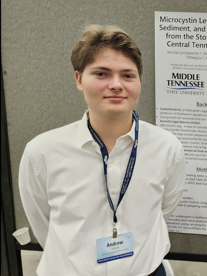
Andrew Hetrick
Seasonal Variations in the Ecology of Harmful Algal Blooms In the Stones River, Tennessee, USA using eDNA and PCR
Faculty Mentor: Dr. Frank Bailey
Harmful Algal Blooms (HABs) are becoming an increasing concern towards water quality and produce toxins that harm people and wildlife. One of the algal toxins of concern, microcystin, is produced by cyanobacteria and acts as a liver toxin. One of the primary bloom-forming genera that produce microcystins is Microcystis spp. The ability of cyanobacteria to produce the toxin depends on the presence of the microcystin synthetase gene cluster within its genome. We used polymerase chain reaction (PCR) techniques on cultured algae to amplify genes that can inform algal community structure in field samples. DNA primers that are capable of detecting genetic sequences for cyanobacteria, Microcystis, and microcystin synthetase E were utilized. Minimum concentrations for detecting bacteria via PCR were determined before use on field samples. These primer sets were then applied to field samples from the East Fork of the Stones River to infer the variation in cyanobacteria communities and toxin-gene presence through time. The Biotechnological data was compared with microcystin concentration, nutrient levels, pH, dissolved oxygen, conductivity, and bioaccumulation in aquatic emergent insects and spiders. The presence of target sequences for Microcystis and microcystin synthetase E were found to show a strong relationship with toxin concentrations. Cultured algal cells will be used again to develop methods that quantify the amount of a gene in a sample for the three primer sets. The relationship between toxin gene presence and microcystin levels indicates that there are future applications of using its abundance in the prediction of harmful algal blooms.

Ariel Nicastro
Improving Zinc Oxide Nanorod Synthesis for Enhanced Electrochemical Sensor Performance
Faculty Mentor: Dr. Suman Neupane
Zinc oxide (ZnO) is a biocompatible inorganic semiconductor with light-emitting and semiconducting properties, making it suitable for diverse applications, including medicine, cosmetics, and nanotechnology. Its wide bandgap and high electron mobility make it particularly promising for advanced sensing technologies. This study focuses on optimizing the hydrothermal synthesis of ZnO nanorods by investigating the effects of autoclave temperature and surfactants. The goal is to produce nanorods with enhanced crystallinity, diameters below 50 nm, and large length-to-diameter ratios—key attributes for improved ZnO-modified sensor performance. X-ray diffraction confirmed the crystalline structure of the synthesized ZnO, while scanning and transmission electron microscopy characterized the nanorod morphology. UV-visible spectroscopy revealed strong absorption at 370 nm, corresponding to a 3.35 eV bandgap, validating the material’s potential for sensor applications. The optimized ZnO nanorods are designed to enhance the sensitivity, accuracy, and scalability of ZnO-modified electrochemical sensors. Sensor performance will be assessed using cyclic voltammetry to track current responses to voltage changes. Future work will refine ZnO integration with electrode materials, explore alternative synthesis methods, and investigate the role of ZnO modification for biosensors and photoelectric sensors. By addressing critical synthesis challenges, this research advances the reliability and cost-effectiveness of ZnO-based sensors, paving the way for their broader use in environmental monitoring, healthcare, and industrial quality control.

Muny Chet
Determining Trophic Movement of Riparian Systems Using Tetragnathid Spiders as a Sentinel Species
Faculty Mentor: Dr. Frank Bailey
The most reported cyanotoxins are hepatotoxins called microcystins. Microcystins are potent hepatotoxins. They are strong inhibitors of the serine/threonine phosphatases which control key biological pathways such as cell proliferation death. This interference triggers a chain of events that leads to eventual death of animal cells. Microcystin targets the liver and induces hepatocyte death. Microcystis blooms that produce microcystin are becoming increasingly more common due to human activity, potentially moving onto terrestrial ecosystems through trophic – food chain – transfer. Tetragnathid spiders’ diets primarily consist of aquatic emergent insects (insects that develop in water before emerging), making them indicators of movement between aquatic and terrestrial ecosystems. Water, sediment, spiders, and insects were collected biweekly at three sites at Walter Hill Dam. All samples, except water, underwent a methanol extraction and analyzed for microcystin concentrations. Previous studies indicated terrestrial transfer of microcystin from mayflies to birds; we expect similar relationships between insects and tetragnathid spiders. Microcystin concentrations were confirmed in aquatic insects and tetragnathid spiders. Considering spiders’ diets are primarily insects, a probable cause of spider microcystin levels being elevated is from consumption of insects. Microcystin concentrations in water, sediment, insect, and spiders were found to change seasonally. Globally, microcystin contamination has caused animal and human deaths. Knowing the potential of it to bioaccumulate can help us understand how to contain it. Future directions for this research will focus more on triggers of microcystin production.
Hannah Antrican
“What Can I Do About It?”: Climate Anxiety in College Students
Faculty Mentor: Dr. Angela Mertig
Climate change presents increasingly worsening global social justice issues. Along with the environmental and economic effects of the changing climate come a variety of personal stressors. Mental health practitioners have noted a growing number of patients expressing distress due to environmental issues. This distress, known as climate anxiety, is a relatively new topic and thus has been used in multiple contexts with varying definitions. It is a topic of relevance to sociologists as well as mental health practitioners. Understanding climate anxiety will aid in promoting action to combat climate change. This research examines the existing literature on climate anxiety, specifically in college-age individuals in preparation for a future study measuring it in the student population of Middle Tennessee State University. Research studying climate anxiety in college students is of particular importance as they represent emerging professionals and scholars who will shape the future of climate policy. Limited research on climate anxiety in college students exists, but a study by the Yale Program on Climate Change Communication showed that Gen Z and Millennials have higher rates of climate anxiety compared to older adults. Existing research has found a negative relationship between climate anxiety and positive mental health in teens and emerging adults. The aim of this research is to provide an overview of research about: (a) ways that climate anxiety has been defined, (b) existing measures of climate anxiety, (c) the relationship between climate anxiety and pro-environmental behavior, and (d) climate anxiety and overall mental wellbeing.

Anna Collins
The Ordinary Price of Zero
Faculty Mentor: Dr. Justin Gardner
The standard economic model is a key player in Shampanier et al.’s Zero is a Special Price: The True Value of Free Products published in 2007. The authors argue that consumer behavior, when presented with a price of zero, or free, is irrational and inconsistent with the standard economic model. This research claims the standard model presented by that work is an inaccurate characterization of consumer theory and that their experimental research design cannot differentiate between the model they presented, and the concept of utility maximization restricted by a budget constraint. Shampanier and colleagues propose a correction to their model that, upon further examination, can easily be accounted for when the research design allows for substitution and income effects. This research demonstrates these effects using mathematical programming to simulate consumer decision-making with various utility functions, such as the Cobb-Douglass functional form. The results show that standard economic theory does an exceptional job of predicting consumer behavior when one good is free. The predictive power of these models relies on their ability to include both income and substitution effects, which are excluded from the “standard” model. Our study shows that Shampainer and colleagues’ model is a special case of standard consumer theory and confirms why the literature has been able to reliably verify their results.

Ariel Nicastro
Modeling Disorder Effects in Quantum Materials
Faculty Mentor: Dr. Hanna Terletska
In condensed matter physics, disorder refers to asymmetries in the crystal structure at the atomic level. There, materials can undergo phase transitions such as the Anderson Localization Transition (ALT), which is responsible for the conductor to insulator transition. Understanding the impact of disorder on quantum materials is critical for material design, as materials’ properties can be manipulated to improve material durability, cost effectiveness, and other desirable characteristics. Such types of improved materials can be used in buildings, airplanes, and machinery. When studying the effects of disorder, it is important to have reliable numerical tools that can properly simulate disorder effects for various classes of quantum materials. To study the effect of disorder on quantum materials, we developed an effective medium quantum cluster method (CDMT) to simulate different types of disorder: binary, box, and Gaussian. These distributions represent different disorder scenarios in technologically important systems, such as alloys and semiconductors. In this project, we will show that our CDMT method captures the experimentally observed impact of disorder on a material’s density of states, i.e. the broadening of the spectrum by disorder. We will also investigate the importance of non-local (beyond single impurity) scattering effects which we model by a finite cluster method.

Connor Prim
Integrating Classical Music Into the Elementary Classroom
Faculty Mentor: Mrs. Leslie Trail and Dr. Kevin Krahenbuhl
Elementary music should expose children to a wide range of musical experiences. Some of those, such as pop music, are easy to incorporate into lessons due to their fun nature but many educators struggle when considering how to effectively teach music from the classical canon. Most students think it is “boring,” and the overwhelmed music teacher might just give in to their whims. However, teachers who think this way are robbing their students of the enrichment these masterworks provide. Still, educators must be strategic about how they implement them in the classroom. Sitting still for an hour while listening to a Brahms symphony might just reinforce students’ negative perceptions about this music. By utilizing movement, mutual orientation, and musical instruments, teachers can get students to engage and relate to classical music in a whole new way, transforming their future and revitalizing the genre for a new generation of listeners to enjoy.
Event Media Coverage
ICUR Student Resources
- Everything You Need to Know Before Applying
- FAQs
- Writing and Submitting Your Abstract
- Developing Your Presentation
- Attending ICUR
- Abstract Rubric
For questions, please contact Madison Yahn at Madison.Yahn@mtsu.edu.

Fall URECA DeadlineThursday, September 3 at 4:30 pm